-
Email info@biomedengoa.org
-
Address 848 N. Rainbow Blvd. #5486 Las Vegas, NV 89107, USA
1Vishwanath Karad MIT World Peace University, Pune, India.
*Corresponding author: Pradnya Siddhivinayak Kulkarni
Vishwanath Karad MIT World Peace University, Pune, India
Email ID: pradnya.kulkarni@mitwpu.edu.in
Received: Feb 13, 2025
Accepted: Mar 12, 2025
Published Online: Mar 19, 2025
Journal: Biomedical Engineering: Open Access
Copyright: Kulkarni PS et al. © All rights are reserved
Citation: Kulkarni PS, Thorat K, Gautam A, Panpalia D, Gole M, et al. Lung disease detection using deep learning algorithms. Biomed Eng Open Access. 2025; 1(1): 1001.
In today’s international, correct and well-timed diagnosis of respiration sicknesses like pneumonia is critical for powerful treatment and advanced affected person consequences. Leveraging deep studying techniques, this research explores diverse methodologies for pneumonia detection from chest X-ray snap shots via dataset augmentation, exploratory statistics evaluation, and transfer getting to know, the study investigates the efficacy of different deep learning architectures, including CNN (89%), DenseNet121 (91%), ResNet50 (79%), and Incaption V3 (88%), in appropriately classifying pneumonia instances. Results indicate promising accuracies, with DenseNet121 rising as the best version. Furthermore, the complete exploration of deep studying methodologies underscores their capability in revolutionizing clinical picture evaluation and disorder detection
Keywords: Transfer learning; CNN; Chest x-ray; Pneumonia; Tuberculosis; COVID-19.
Motivation
In the realm state-of-the-art scientific imaging, the correct diagnosis latest pneumonia, a respiratory infection affecting hundreds of thousands worldwide, remains a essential undertaking [6]. Pneumonia claims the lives cutting-edge approximately seven-hundred,000 kids yearly and aflicts 7% contemporary the worldwide population [1]. Conventional analysis relies heavily on chest X-rays, but the nuanced interpretation of these snap shots poses problems, even for educated radiologists [9].
To address this pressing difficulty, modern methods leveraging deep today’s fashions have emerged, aiming to enhance diagnostic accuracy and aid clinical professionals in selection making. This paper affords a comprehensive exploration cutting-edge various deep state-of-the-art methodologies applied to pneumonia detection from chest X-ray pics. The studies landscape contains a myriad state-of-the-art study utilizing diverse deep brand-new architectures, statistics augmentation strategies, and transfer latest strategies [4].
From the inception modern-day Convolutional Neural Networks (CNNs) to the modern-day improvements in version architectures consisting of VGG16, ResNet, Inception, and Inception, the quest for an foremost solution maintains [14].
Research contributions
One distinguished technique proposed in this observe involves the improvement today’s an ensemble version leveraging deep today’s architectures like ResNet18, Inception, InceptionV3, DenseNet121, and MobileNetV3 [8]. This weighted classifier amalgamates predictions from character fashions, showcasing advanced overall performance in pneumonia detection compared to standalone models [11]. Transfer ultramodern, excellent-tuning, and partial records augmentation techniques improve the training technique, improving version robustness and generalization [3].
Moreover, the paper explores the efficacy modern CNN models educated from scratch, emphasizing the importance modern day characteristic extraction and class in pneumonia diagnosis [13]. Leveraging geometric augmentation and facts augmentation algorithms, those fashions attain notable validation accuracy, contributing to the developing frame modern literature on deep cutting-edge in medical imaging [2].
Moreover, the look at delves into the utility brand new superior deep learning techniques in the context state-of-the-art COVID-19 prognosis, highlighting the urgency modern leveraging chest X-ray scans for fast and fee-powerful identification present day the virus [15]. Through using updated VGG16 models and innovative photo cropping strategies, the research achieves impressive accuracies in classifying COVID-19, normal, and pneumonia cases [7].
Moreover, the paper provides novel techniques for tuberculosis diagnosis, lung nodule detection, and activity assessment present day pulmonary tuberculosis, the usage of ensemble CNN architectures and revolutionary preprocessing algorithms [12]. These improvements underscore the ability of modernday deep modern day in revolutionizing sickness prognosis and remedy across diverse scientific domains.
Literature review
This study demonstrates the efficacy of deep transfer studying techniques in automated pneumonia detection from chest X-ray pictures. Through the usage of complicated but computationally efficient deep networks, the proposed technique addresses the mission of constrained get admission to radiology diagnostics, mainly in areas with sparse expert assets [1]. Switch mastering and records augmentation mitigate overfitting, at the same time as a weighted classifier efficaciously combines exceptional architectures. The executed excessive accuracy, bear in mind, precision, and AUC score validate the robustness of the model, with capacity implications for improving pneumonia analysis globally. Future studies should explore methods for more green weight estimation and comprise patient records for superior predictions.
This Literature highlights the development of a Convolutional Neural Network (CNN) version educated from scratch for pneumonia detection in chest X-ray photographs via eschewing reliance on transfer gaining knowledge of, the have a look at addresses challenges in reliability and interpretability common in scientific imagery evaluation [2]. Leveraging statistics augmentation techniques, the CNN model achieves terrific validation accuracy, distinguishing among superb and poor pneumonia instances. Destiny extension’s purpose is to tackle the type of X-ray pics containing both lung cancer and pneumonia, addressing an extensive difficulty in clinical imaging.
This study demonstrates the development of a deep studying framework for pneumonia type, presenting four CNN fashions such as ResNet152V2 and MobileNetV2, with fashions designed from scratch [3]. Through rigorous assessment towards recent studies, the proposed framework achieves great improvements in accuracy, precision, F1-score, consider, and AUC. Appreciably, the ResNet152V2 model exhibits superior performance in comparison to other current works, while the MobileNetV2, CNN, and LSTM-CNN fashions additionally showcase marvelous effects, exceeding 91% in key metrics, those findings underscore the efficacy of the proposed deep learning method in improving pneumonia detection and type.
This Literature showcases a novel approach for pneumonia detection using a deep neural network architecture, emphasizing the combination modern day dropout inside the convolutional layers [4]. In contrast to conventional methods reliant on switch getting to know modern, the proposed CNN model is constructed from scratch, presenting superior accuracy and performance in pneumonia detection from X-ray photos. Examined on a dataset from Kaggle’s scientific imaging mission, the version achieves superb metrics, inclusive of 97.2% accuracy, 97.3% consider, and 97.4% precision, rivaling solutions. Considerably, the proposed model demonstrates superior overall performance and rapid predictions, with remember and precision exceeding 97% and inference time as little as 122 ms.
This literature highlights the urgency of pneumonia detection and the pivotal position of deep gaining knowledge of strategies in scientific imaging. With a baby succumbing to pneumonia every 39 seconds, early identity becomes imperative [5]. At the same time as chest X-rays are common diagnostic gear, distinguishing pneumonia from other lung situations affords a mission. Leveraging deep learning, particularly ConvoLutional Neural Networks (CNNs), offers a promising solution. Pre-trained CNN models serve as effective feature extractors for classifying abnormal chest X-rays. Amid the COVID-19 pandemic, CNNs were instrumental in detecting the virus from X-ray pictures. In this have a look at, various deep studying models together with ANN, CNN, and VGG19 were hired, yielding widespread detection accuracies.
This study demonstrates the pressing want for accurate pneumonia diagnosis using superior gadget modern day techniques in medical imaging. Leveraging a singular multiclass machine ultra-modern framework, the B2-internet version, researchers reap excellent accuracy, keep in mind, and AUROC scores, distinguishing among every day, bacterial, and viral pneumonia in chest X-ray photographs. Through retraining pinnacle-acting fashions and using ensemble strategies, the B2-internet framework outperforms current tactics, showcasing its capacity for scientific implementation. The look underscores the importance of trendy AI and ML in healthcare selection-making, highlighting the importance of modern green diagnostic models to enhance affected person results [6].
This Literature highlights the pressing need for green COVID-19 diagnosis strategies amidst the worldwide pandemic, leveraging chest X-ray scans and deep learning strategies [7]. Employing picture cropping and an up-to-date VGG16 version, the take a look at achieves astounding type accuracy across 3 public datasets, notably, the version reduces parameter complexity even as keeping high overall performance, exhibiting 99% accuracy in COVID-19 category. The studies outline a scientific approach, inclusive of information preprocessing, version schooling, and overall performance evaluation, contributing to the advancement of speedy and price-effective diagnostic answers. Destiny endeavor’s goal is to extend this system to various medical imaging domain names, promising broader packages in sickness class and healthcare choicemaking.
This study demonstrates the ability of deep studying-primarily based CNN fashions in classifying Covid-19 infected patients the use of chest X-ray scans, imparting valuable insights for fast screening amidst the pandemic [8]. Through experimentation with a couple of CNN architectures, which include Inception V3, Inception, and ResNet, the Inception model emerges because it is the maximum accurate, achieving 97.9% accuracy. At the same time as acknowledging the want for in addition validation against new datasets and session with clinical specialists, the research underscores the feasibility of leveraging deep gaining knowledge for automatic prognosis responsibilities in preventing Covid-19. This work emphasizes the significance of economically possible solutions in addressing the demanding situations posed with the aid of the pandemic, paving the manner for destiny research endeavors [8].
This study introduces an innovative ensemble method, SGDRE, based on stochastic gradient descent with warm restarts, addressing key challenges in childhood pneumonia identification from chest X-ray images [9]. Leveraging the strengths of ensemble methods and SGDR, SGDRE achieves superior generalization capabilities while mitigating the demand for large datasets and reducing time complexity. Trained on pediatric chest X-ray images, SGDRE exhibits compelling performance, surpassing baseline methods with a test accuracy of 96.26% and AUC of 95.15%. The method’s ability to navigate multiple minima and generate diverse models within a single training process underscores its efficacy in enhancing classification accuracy for childhood pneumonia diagnosis.
This literature implements a singular technique utilizing Convolutional Neural Community (CNN) architecture estimation via Bayesian optimization for the detection and classification of pneumonia, distinguishing between viral and bacterial types. With pneumonia being a main motive of mortality, fast and correct diagnosis is paramount. The proposed approach achieves promising effects, boasting a 0.964 accuracy for pneumonia detection and a 0.957 accuracy for pneumonia type category, without employing not unusual preprocessing strategies like histogram equalization and lung segmentation. This underscores the efficiency and effectiveness of the proposed method in developing high-overall performance neural networks for pneumonia analysis, imparting capability improvements in scientific decision-making.
This study implements a novel approach using Convolutional Neural Networks (CNNs) for automating the differentiation between COVID-19-inflamed individuals and wholesome ones using chest CT scans [11]. With the aid of using binary classifications as opposed to an unmarried multiclass class, the proposed version minimizes fake predictions, enhancing accuracy, sensitivity, and F1-rankings important for contamination manipulation. The model offers fast prediction, making it fee-powerful in comparison to RT-PCR assessments, albeit not ready for production. Whilst the version indicates promising accuracy, it’s encouraged for studies functions, with confirmation from a clinician or radiologist advised. This painting gives valuable insights for future studies and alertness in COVID-19 prognosis.
This paper implements three proposals utilizing pre-trained CNNs for tuberculosis detection, showcasing aggressive effects compared to present literature. Notion 1 employs CNN architectures to extract functions from radiographic photographs for SVM classification. concept 2 extracts feature from subregions of the chest radiograph, improving type thru combined worldwide descriptors. Notion 3 integrates SVMs from Proposals 1 and a pair of into classifier ensembles. Notwithstanding pre-educated networks’ inferiority to task-precise models, they provide efficiency and effectiveness, especially in high-decision datasets. Destiny advancements in tuberculosis detection may hinge on the creation of large annotated datasets, akin to the ones seen in dermatology, to reinforce CAD machine efficacy [12].
Dataset description
The data description elucidates the procedure of acquiring the dataset from Kaggle, comprising images classified into normal and pneumonia lessons, dispensed across education, trying out, and validation folders [7]. The following conversion of pixels into NumPy arrays helps standardization and performance in data processing, an essential step in making ready the dataset for model education and evaluation. The depiction of picture distribution over binary instructions, aided through visualization strategies, offers insights into dataset composition and ability class imbalances, guiding next information dealing with techniques [4].
Dataset description
Statistics augmentation techniques play a pivotal position in improving the robustness of deep studying fashions. The application of normalization techniques ensures consistency in pixel values throughout pics, facilitating convergence at some point of model education [10]. Reshaping of image arrays into suitable tensor codecs enables compatibility with deep mastering architectures, a prerequisite for effective version implementation [2]. Augmentation strategies, consisting of rotation, zooming, and moving, introduce versions in training facts, mitigating overfitting and enhancing model generalization. These augmentation strategies enhance the schooling dataset, enhancing the variety of representations and thereby improving version performance.
Exploratory data analysis (EDA)
Exploratory facts analysis (EDA) serves as a foundational step in information dataset characteristics and distribution styles [5]. Via visualizations such as count plots and pattern picture shows, researchers benefit from insights into class distribution, photograph exceptional, and dataset diversity [11]. The identification of ability demanding situations, inclusive of class imbalances or information exceptional problems, informs next information preprocessing and version improvement decisions. Common, EDA presents a complete evaluation of the dataset, facilitating knowledgeable picks in model design and schooling strategies [5].
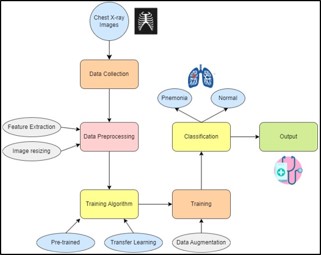
System architecture
The system structure depicted in Figure 1, illustrates the workflow of Tuberculosis (TB) and pneumonia detection methodologies. At the center of the structure are three class methodologies: Random woodland, Deep CNN, and voting classifiers, every tasked with scrutinizing present research [11]. Thru a thorough exam, our observe targets to discover gaps in research, such as version balance, magnificence imbalance, and segmentation demanding situations [8]. A complete literature evaluate will delve into factors influencing TB and pneumonia detection effects, consisting of dataset selection standards and evaluation metrics [9]. Drawing insights from this evaluation, our research will suggest destiny directions for enhancing detection methodologies [12]. Leveraging advancements in gadget mastering and clinical imaging generation, our have a look at ambitions to deal with identified gaps and contribute to advanced TB and pneumonia detection methods [10]. ultimately, this systematic technique seeks to decorate patient effects by enabling extra correct and timely prognosis of TB and pneumonia [6].
Experimental results
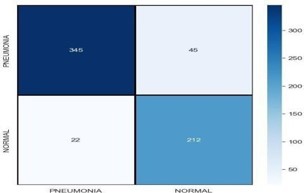
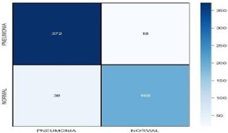
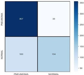
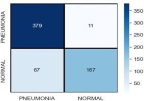
A confusion matrix is a overall performance dimension tool used in device gaining knowledge of to assess the accuracy of a class model [2]. It’s far in particular beneficial when operating with binary class problems, despite the fact that it may also be extended to multi-class class. Classification of images into Pneumonia and normal were tried using various machine learning models such as Convolution Neural Network, Densenet121, Resnet 50 and Inception. Figures 2,3,4,5 shows confusion matrix after testing the various models.
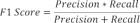
F1-Score is the harmonic mean of precision and recall. It is suited for imbalanced datasets. The formula is as shown in Equation (7).
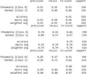
Following are classification reports of all the models from Figure 6
Model assessment includes evaluating the overall performance of various gadget mastering models primarily based on their accuracies in classifying statistics. This contrast is generally visualized the usage of a bar graph, in which every version is represented with the aid of a bar whose peak corresponds to its accuracy rating [7]. All model accuracies are compared, by way of examining these bars, researchers can effortlessly perceive which version plays pleasant overall. Additionally, they could examine the relative strengths and weaknesses of every version based totally on their accuracy scores. This visualization aids in choice-making regarding the choice of the maximum appropriate version for a selected project or dataset [4]. The comparison is shown in Table 1 and a short comparison of accuracies of other paper’s accuracies is done in Table 2.
| Model | Accuracies |
|---|---|
| CNN | 89% |
| DenseNet121 | 91% |
| ResNet50 | 79% |
| InceptionV3 | 88% |
| Ref No. | Model | Test Accuracies |
|---|---|---|
| [ours] | DenseNet121 | 91% |
| [3] | ResNet152V2 | 91% |
| [4] | CNN | 97.2% |
| [7] | VGG16 | 99% |
| [8] | Inception V3 | 97.9% |
| [9] | SGDRE | 96.26% |
| [10] | CNN | 95.7% |
Out of all the models applied, DenseNet emerged as the fine-performing model in terms of testing accuracy. To evaluate its efficacy, a detailed evaluation turned into conducted on each successfully and incorrectly anticipated photographs. Efficaciously predicted photographs exhibit instances wherein the version accurately classified the records in line with the floor fact. Conversely, examining incorrectly anticipated pix sheds light at the model’s obstacles and regions for improvement. This qualitative assessment process enables in understanding the version’s behavior, identifying capacity assets of errors, and refining the model for better overall performance in destiny iterations [5]. Collaborative efforts among researchers, clinicians, and technologists will play a critical position in driving innovation and ultimately improving patient care by presenting quicker, more correct diagnostic tools available to all.
The preceding discussions underscore the burgeoning potential and diverse applications of deep learning, particularly in medical image analysis, from pneumonia and COVID-19 detection to tuberculosis diagnosis. Future advancements may involve refining existing methodologies, such as ensemble learning and pre-trained CNNs, to enhance accuracy and efficiency further. Moreover, expanding datasets and integrating emerging technologies like reinforcement learning and attention mechanisms could revolutionize disease detection and classification. Collaborative efforts between researchers, clinicians, and technologists will be pivotal in navigating these frontiers, ultimately advancing healthcare by providing faster, more accurate diagnostic tools accessible to all.